I know this is a long write up, the first half is biochemistry and what happens on a cellular level. The second half is more pertaining to the average AAS user, including a deeper dive into liver functioning tests and liver support.
I highly recommend at least reading the second half, especially the Liver Support section. Hepatotoxicity is a word that is frequently thrown around, everyone’s heard it, everyone thinks they know what it is, but once you ask something beyond surface level, you get a whole lot of conflicting answers. Let’s dive into it.
Overview/Background/General Information/What the fuck actually happens? Drug Metabolism: The human body identifies almost all drugs as foreign substances and subjects them to various chemical processes to make them suitable for elimination. Drug metabolism is typically split into two phases: Phase 1 (oxidation via Cytochrome P450, reduction, and hydrolysis) tends to increase water solubility of the drug and can generate metabolites. Phase 2 further increases water solubility of the drug, inactivating metabolites, thus preparing it for excretion.
- Aside: There has been a lot of questions regarding using high dose psoralens (from grapefruit juice/extract) to inhibit the activity of Cyp3A4 (and the Cyp450 family in general) or consuming large quantities of chargrilled meat to induce the activity Cyp450. DO NOT purposely do this in an attempt to increase the bioavailability of oral steroids. The Cyp450 family is incredibly important enzyme family that is involved in the metabolism (both activation and degradation depending) of statins, oral contraceptives, acetaminophen, anti-depressants, beta-blockers, antiarrhythmic agents, and many, many more. This can cause SEVERE adverse effects.
- Example: Acetaminophen (Tylenol) is often seen as hepatotoxic, it is not. One of the metabolites, NAPQI, is. Under normal conditions, NAPQI is produced in incredibly minor amounts. Alcohol induces Cyp450 enzyme family, which increases the breakdown of Tylenol into NAPQI, causing intensive acute liver damage which can often be fatal in high doses. In short, don’t nurse your hangover with Tylenol, use Advil instead… or better of just don’t drink alcohol.
17α-Alkylated Anabolic Steroids. These AAS contain a methyl or ethyl group on the C17α position, allowing for oral activation. This modification allows the drug to survive hepatic metabolism, limiting the amount of steroid that is broken down, allowing for more drug to reach the bloodstream. Without this modification, the drug is completely broken down by the liver, never reaching systemic circulation. This initial process is called First Pass Metabolism.
First pass metabolism: After a drug is swallowed, it is absorbed by the digestive system and enters the hepatic portal system. It is carried through the portal vein into the liver before it reaches the rest of the body. The liver metabolizes many drugs, sometimes to such an extent that only a small amount of active drug emerges from the liver to the rest of the circulatory system. This first pass through the liver may greatly reduce the bioavailability of the drug. Some oral steroids have a very low bioavailability due to first pass metabolism, thus injectable versions may be used to prevent the initial breakdown, effectively increase bioavailability and reducing liver stress.
- Aside: There have been questions regarding sublingual administration of oral AAS in order to bypass first pass metabolism. In short, very few drugs can be properly absorbed sublingually, Nitroglycerin is a prime example. AAS, although can be manufactured for sublingual absorption it requires a specific method of delivery (sublingual tablets, strips, or sprays for example), which are difficult to obtain and manufacture; it’s not as simple as placing some powder under your tongue. Other drawbacks exist of sublingual administration such as tooth decay and difficulty in dose management.
- Anavar: The exception to the rule: The oral bioavailability of oxandrolone is 97%. The drug is metabolized primarily by the kidneys and to a lesser extent by the liver. Oxandrolone is the only AAS that is not primarily or extensively metabolized by the liver, and this is thought to be related to its diminished hepatotoxicity relative to other AAS. For this reason, there is no reason to ever use injectable anavar (unless poking yourself that often is your kink, no judgement).
In short: Oral Steroid (active) -> Hepatic Breakdown -> Metabolite (inactive) - [aside: not all drugs are orally active, in fact the majority are inactive. For example, codeine (inactive) will be converted into the active form, Morphine, within the liver via Cyp450 enzymes]
In the case of oral AAS, hepatic metabolism can convert an active drug into its inactive form; C17α methylation prevents this. Why is this modification known to be hepatotoxic? The
primary enzyme that normally breaks down hormonal steroids (such as endogenous DHEA, testosterone, estradiol, etc) is 17β-Hydroxysteroid dehydrogenase, 17β-HSD, (and to a minor extent the Cyp450 family) which can no longer break down the methylated drug, thus the liver finds an alternative route for metabolism. The actual specific process is still relatively unknown, but involves a variety of oxidation reactions, inducing an increase of free oxygen radicals within the hepatocytes (liver cells), causing cell death due to oxidative stress.
There is another hypothesis which involves the presence of androgen receptors within the liver. The C17α methylated oral steroid, that is no longer properly broken down, will bind to these receptors, causing a drastic increase of androgenic activity within the liver, leading to hepatoxicity.
In my opinion, it is a mixture of both. Many studies show a direct correlation between the androgenic effect of the oral steroid and the amount of hepatoxicity. The exact link between the two is yet to be determined.
In general, the greater the affinity of C17α methylated oral steroid for the androgen receptor, the more hepatoxicity occurs. Hepatotoxicity is an overlying term: the specifics related to AAS use are Cholestasis (blockage of biliary flow), Steatosis (accumulation of fatty lipids within the liver), Zonal Necrosis (hepatocyte death within a specific zone of the liver), and Peliosis Hepatitis (vascular lesions leading to liver enlargement).
- Aside: Steatosis (fatty liver) has been an observed adverse effect of Nolvadex and Raloxifene
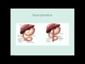
Cholestasis is a condition where bile cannot flow from the liver to the duodenum. It is the most common condition resulting from oral AAS use. In short, bile is continuously produced but cannot leave the liver, causing build up, backflow, and eventually hepatocyte death. Differential symptoms of cholestasis include but are not limited to pruritus (itchiness), jaundice (yellowing of the skin and whites of the eyes), pale stool, and dark urine.
Liver Functioning Tests: What do they mean and why are they relevant?
AST: Aspartate Transaminase: This alone is not a good indication of liver damage. AST is found in abundance within both cardiac and skeletal muscle. An elevated AST value can be caused by something as minor as weightlifting.
ALT: Alanine Transaminase: ALT is found specifically within the liver and is released into the plasma when significant liver stress, including hepatocyte death, occurs. An elevated value is of concern.
- Aside: general rule of thumb (not always true) if both elevated are elevated and if AST>ALT in a ratio of 2:1, suspect alcohol/drug induced damage. If both values are elevated and if ALT>AST in a ratio of 2:1, suspect viral hepatitis.
ALP: Alkaline Phosphatase: ALP is found within the hepatobiliary ducts. An elevated value is commonly indicative of obstruction and bile buildup, signifying cholestasis.
GGT: Gamma-glutamyl Transferase: GGT is an enzyme that is found in many organs throughout the body, with the highest concentrations found in the liver. GGT is elevated in the blood in most diseases that cause damage to the liver or bile ducts.
5’-nucleotidase: The concentration of 5’-nucleotidase protein in the blood is often used as a liver function test in individuals that show signs of liver problems. ALP can be elevated due to both skeletal disorders and hepatic disorders. 5’-nucleosidase is elevated ONLY with hepatic stress, not skeletal, thus allowing for differentiation.
Putting it all together: Cholestasis can be suspected when there is an elevation of both 5'-nucleotidase and ALP enzymes. Normally GGT and ALP are anchored to membranes of hepatocytes and are released in small amounts in hepatocellular damage. In cholestasis, synthesis of these enzymes is induced, and they are made soluble. GGT is elevated because it leaks out from the bile duct cells due to pressure from inside bile ducts. As hepatocyte damage continues, ALT, AST, and unconjugated bilirubin will begin to rise.
In short: Initial liver stress causes 5’-nucleiotidase and ALP to rise, shortly after GGT rises, then finally AST and ALT rise. Thus, with only AST and ALT values, it is difficult to determine the cause and extent of hepatic damage.
Liver Support: NAC/TUDCA/Liv52 NAC: N-Acetyl Cystein NAC is a prodrug of L-cysteine, a precursor of the biological antioxidant glutathione which is able to reduce free radicals within the body. Free radicals, which as discussed above, are associated with causing extensive hepatocyte damage due to the oxidative breakdown of C17α methylated AAS.
In addition to its antioxidant action, NAC acts as a vasodilator by facilitating the production and action of nitric oxide. This property is an important mechanism of action in the prophylaxis of contrast-induced nephropathy and the potentiation of nitrate-induced vasodilation.
- Aside: NAC has been constantly used as an adjunct to multiple neurological disorders including Parkinson’s, Huntington’s, Multiple Sclerosis, Cerebral Ischemia, Alzheimer’s and MANY more due to the potent free radical trapping effect which prevent mitochondrial dysfunction.
Multiple studies have constantly showed NAC decreasing liver functioning tests and improving liver function and mitigating cholestasis. NAC had the ability to vastly improve markers of kidney function and was actually able to even double the rate of sodium excretion, indicating that NAC is may be useful in preventing water retention.
In short, NAC has a vast number of benefits, including hepatoprotective (liver), nephroprotective (kidney), and neuroprotective (neural), and anti-inflammatory effects that have been constantly demonstrated thru literature. Moreover, NAC can and should be used for year-round support since the adverse effects are incredibly mild. There is absolutely NO reason to not be taking NAC.
TUDCA: Tauroursodeoxycholic acid TUDCA is a bile acid taurine conjugate form of UDCA. As discussed above, during cholestasis, bile builds up, creating backflow and inducing hepatocyte death thru apoptosis. Apoptosis, or programmed cell death, is largely influenced by the mitochondria. If the mitochondria are distressed, they release the molecule cytochrome C. Cytochrome C initiates enzymes called caspases to propagate a cascade of cellular mechanisms to cause apoptosis.
TUDCA prevents apoptosis with its role in the BAX pathway. BAX, a molecule that is translocated to the mitochondria to release cytochrome C, initiates the cellular pathway of apoptosis. TUDCA prevents BAX from being transported to the mitochondria, effectively inhibiting hepatocyte death.
Furthermore, TUDCA aids in the processing of toxic bile acids into less toxic forms, resulting in decreased liver stress, further preventing hepatocyte death. Moreover, TUDCA aids in the transport of bile from the liver into the duodenum, effectively unblocking the build up causing cholestasis. Finally, TUDCA has been proven to be an effective treatment for the necro-inflammatory effects of Hepatitis. Study after study has shown that TUDCA greatly improves liver enzyme values.
Why do we recommend only using TUDCA with hepatotoxic oral steroids? The idea is that TUDCA induces liver damage when there is no hepatotoxicity present… but after reading the above, does that make sense? It does not.
I could not find any literature showing that TUDCA induces liver toxicity. The recommendation instead is due to the negative effects of TUDCA on cholesterol values. TUDCA has been shown to greatly decrease HDL levels when taken for extended periods of time. The idea is, if you have a healthy functioning liver, there is no reason to take TUDCA for long periods of time since all you’re doing is decreasing HDL values. That being said, after doing the research and seeing the vast benefits of TUDCA (included bellow, not a comprehensive list), I am beginning to change my perspective on TUDCA use with only hepatotoxic oral AAS.
In short, TUDCA prevents hepatocyte death, enhances hepatocyte function, exhibits anti-inflammatory effects on the liver, neutralizes toxic bile, and prevents bile build up that was caused by the alternative metabolism of C17α methylated AAS. ***THERE IS NO EVIDENCE THAT I HAVE COME ACROSS THAT SHOWS THAT TUDCA ITSELF INDUCES LIVER DAMAGE WHEN USED WITHOUT HEPATOTOXIC DRUGS**\*
TUDCA has a variety of other benefits outside the liver, but I will not go into them this time. In short: - Increasing glucose-induced insulin release via the cAMP/PKA pathway, increasing insulin sensitivity.
- Relieving endoplasmic reticulum (ER) stress. The ER makes sure proteins are folded properly.
- Reducing programmed cell death (apoptosis) in healthy cells. TUDCA prevents the molecule BAX from reaching the mitochondria. BAX causes mitochondria to release cytochrome C, which causes enzymes (caspases) to initiate apoptosis.
- Inactivating Bcl-2-associated death promoter (BAD), a molecule involved in apoptosis.
- Removing toxic bile acids from the liver and preventing them from damaging liver cells
- Exhibits neuroprotective effects via the inhibition of NF-kB
- Preserve photoreceptor function and increases retinal health by increasing rd10
Sources
Liv52 Liv52 is an herbal liver support. There have been medical studies conducted on Liv.52 in recent years, many of which involve its ability to protect the liver from damage by alcohol or other toxins. Liv52 has been shown to exhibit antiperoxidative function, antioxidant effects, anti-inflammatory, diuretic effects and neutralization of toxic products within the liver.
“The results demonstrated that the patients treated with Liv-52 for 6 months had significantly better child-pugh score, decreased ascites, decreased serum ALT and AST. We conclude that Liv-52 possess hepatoprotective effect in cirrhotic patients. This protective effect of Liv-52 can be attributed to the diuretic, anti-inflammatory, anti-oxidative, and immunomodulating properties of the component herbs.”
“Liv.52 enhanced the rate of absorption of ethanol and rapidly reduced acetaldehyde levels, which may explain its hepatoprotective effect on ethanol-induced liver damage.”
“Liv.52 administration reduced the deleterious effects of ethanol. The concentration of acetaldehyde in the amniotic fluid of ethanol-consuming animals was 0.727 microgram/ml. Liv.52 administration lowered it to 0.244 microgram/ml. The protective effect of Liv.52 could be due to the rapid elimination of acetaldehyde.”
That being said, there is conflicting research on Liv52. The studies either show hepatoprotective function or no effect, positive or negative. “There was no significant difference in clinical outcome and liver chemistry between the two groups at any time point. There were no reports of adverse effects attributable to the drug. Our results suggest that Liv.52 may not be useful in the management of patients with alcohol induced liver disease.”
In short, Liv52 can be used if you have the additional funds, it is not the end-all-be-all but can be used as an adjunct. It is an incredibly cheap drug that may improve liver function and exhibit hepatoprotective effects. IT SHOULD IN NO WAY YOUR ONLY LIVER SUPPORT MEDICATION, but there is nothing wrong with using it. As mentioned in the syllabus one aspect of Biochemistry deals with two direct yet deceptively simple questions. These are: (1) where did it come from, and (2) where does it end up?
In this lecture, we will ask these two questions to the hemoglobin-oxygen complex. As we answer these questions, we will touch upon oxygen transport to the cells, the expression and degradation of proteins, and the biosynthesis and breakdown of heme.
In the introductory
lecture, we attempted to perform some run-of-the-mill, double-entry accounting. Each time we inhaled, we learned that a certain percentage of oxygen molecules were getting ‘fixed’ inside our body. Let’s pick up from there.
The oxygen we inhale not only dissolves in our blood, but it also binds tightly to a special protein found in our erythrocytes called Hemoglobin. The protein data bank (PDB) contains more than 80,000 structures of proteins, and the following
GIF was downloaded from that site. The PDB also has a section called the molecule of the month. In this section, they describe in crisp, atomic detail how some select proteins do what they do. Their section of hemoglobin is linked
HERE. Specifically, one molecule of oxygen binds to a central Iron atom in another molecule called heme. The heme molecule contains 4-pyrrole rings linked by methylenes. Hemoglobin, contains a heme portion, and a protein portion called globin. Oxygen is transported to the tissues by means of this oxygen-hemoglobin complex. Let us pay close attention to the structure of this complex and ask: (1) where did it come from, and (2) where does it end up?
In the case of this particular complex, let us divide the first part into 3 subsections, and ask: (a) where did the oxygen molecule come from, (b) where did the globin protein come from, and (c) where did the heme ring come from? Similarly, we can divide the second question into 3 subsections: (a) where will the oxygen end up, (b) where will the globulin protein end up, and (c) where will the heme ring end up? Let’s address each of these questions one by one:
Oxygen 1 (a): Where did the oxygen come from?
- Oxygen is present in the air we inhale, and most of the oxygen molecules in air come from plants. Specifically, the oxygen molecules come from deep inside a plant’s chloroplast. There, each oxygen molecule is created from two molecules of water by a protein-complex known as the oxygen-generating complex. Inside this complex, the oxygen atoms of two water molecules get hitched together into one oxygen molecule. Prior to this moment, these two water molecules were independent. They may have originated from two very different places. However, after a trip to the chloroplasts, the oxygen atoms of these two water molecules get hitched, and now they must spend their lives together as one oxygen molecule. (Imagine the first water molecule evaporating from the surface of the Pacific Ocean, near Hawaii, 10 months ago; and the second water molecule evaporating from the surface of the Indian Ocean, near the South African coast, two years ago. The two water molecules, then, floated as clouds, until last week when they drizzled-down as rain on our neighborhood park. After working their way through the wet mud, and then up some plant root, they finally reached the oxygen-generating complex of a blade of grass, together. On this blade of grass, the oxygen atoms of these two, formerly independent, water molecules, that originated from two random places on the planet, were hitched together to produce one oxygen molecule. This oxygen molecule was floating in the air until just a few seconds ago. Then, it entered our lungs and was promptly picked up by our hemoglobin).
2 (a): Where will the oxygen end up?
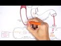
- This answer also requires a story. In the lungs, oxygen molecules do two things: (1) they dissolve into the blood solution, and (2) they bind to hemoglobin. Hemoglobin behaves as an oxygen reserve. If we only relied on the ability of oxygen to dissolve in blood, we would have nowhere near all the oxygen our body needs. The ability of hemoglobin to bind an oxygen molecule, and subsequently release it, allows us to achieve ~70 fold higher concentrations of oxygen. Nitrogen molecules don’t bind hemoglobin very well, but they do dissolve in blood. Carbon monoxide, on the other hand, binds hemoglobin ~200 fold more tightly than oxygen. If carbon monoxide was present in the inhaled air mixture, it would out-compete oxygen for binding to hemoglobin. Carbon monoxide thus leads to some pretty serious problems. Coming back to the complex of oxygen and hemoglobin, we notice that the complex is itself enclosed inside erythrocytes. As the heart beats, blood is pumped through the body, and the erythrocytes that were near the lungs now start flowing away. As these cells flow away, the oxygen molecules start leaving the oxygen-hemoglobin complex, and enter into the blood solution, from where they enter into other cells in the body. In the muscle, oxygen molecules can also bind to myoglobin, a protein like hemoglobin. Eventually, most oxygen molecules find themselves at the edge of the mitochondrial inner membrane, deep inside cells. Up to this stage, the entire transport process, or the journey, of oxygen from our lungs to the edge of the mitochondria can be understood more or less as a physical process. In other words, it can be understood as a series of binding and unbinding events between oxygen molecules, proteins, and the blood. What happens next to the oxygen molecules at the edge of the mitochondrial inner membrane is not a physical transformation but a chemical one. The transformation of oxygen to water will be a topic for discussion in Lecture 2.
Globin 1(b): Where did the globin come from?
- The globin protein was synthesized from DNA by using several different RNA: including ribosomal RNA, transfer RNA, and messenger RNA. Considerable amount of energy was spent to do this. In fact, protein synthesis is one of the most expensive things a cell can do. The information in the globin gene (which is structurally represented by a strand of DNA) was transcribed on to messenger RNA, and then this protein was synthesized, one expensive peptide bond at a time.
2(b): What will happen to the globin?
- Most erythrocytes have a life-span of ~3 months. As the RBC die, the globin protein is broken down to its individual amino acids. Amino acids are considered too valuable to be squandered as fuel by the body, so they are typically re-used. However, under some circumstances, amino acids can also be burned as fuel by involving transamination, the Urea cycle and the TCA cycle.
Heme 1(c): Where did the heme ring come from?
- The heme ring was synthesized in our body by a series of enzymatic transformations from the amino acid glycine and succinyl CoA, a molecule of the TCA cycle. (The TCA cycle is also a topic for later discussion). First, glycine combined with succinyl CoA to generate a product that upon decarboxylation gave δ-Aminolevulinic acid (ALA). Two ALA molecules were then condensed to give porphobilinogen. Subsequently, four porphobilinogens were deaminated give hydroxymethylbilane that cyclized to uroporphyrinogen. The uroporphyrinogen was decarboxylated to coproporphyrinogen, which was subsequently oxidized to protoporphyrin. Heme is obtained once iron adds to protoporphyrin. The biosynthesis of heme is important because we all need heme to bind oxygen. However, another reason why these transformations are important is because their malfunction results in disease. Specifically, improper execution of some of the steps of heme biosynthesis leads to porphyrias (diseases in which the urine exhibits unnatural colors).
2(c): Where will the heme end up?
- When erythrocytes die, the iron atom is stripped-off from heme and recycled. The protoporhyrin is converted into biliverdin, which is then transformed to bilirubin. Bilirubin is not very soluble, and must be conjugated with glucuronic acid to make it more soluble. This conjugate exits the body via the bile. In addition, intestinal bacteria can convert a small portion of conjugated bilirubin to urobilinogen – a molecule excreted in the urine, and a larger portion of conjugated bilirubin into stercobilinogen - a molecule excreted in the stool.
Let’s step back, and take a moment and think about the origin and fate of biomolecules. For the past three months a heme molecule, bound to globin, dutifully brought oxygen from all over the planet, to every cell in our body. It brought oxygen to the muscles of our heart, allowing it to beat. It brought oxygen to the neurons in our brain, contributing to our thoughts. However, very soon this heme molecule will leave our body. Right now, there might also be some spanking new heme molecules binding to oxygen for the first time in their life. And then, in around three months, they too will be out of our body.
This is also a good place to introduce the winner of the 1930 Nobel Prize in Chemistry: Hans Fischer and the
Award Speech Assignment 1: Draw the following structures showing interatomic bonds as lines: You can use a white board and marker, pen/pencil and paper, or use any freely downloadable drawing software. - (1) Oxygen
- (2) Water
- (3) Carbon Monoxide
- (4) Pyrrole
- (5) Glycine
- (6) Succinic Acid and Succinate
- (7) Glucuronic Acid
- (8) Heme
Assignment 2: Write a short research memo (one or two paragraphs) about any Porphyria using available internet resources. Bilirubin comes from the breakdown of red blood cells and is excreted by the liver. High levels of bilirubin can cause jaundice, which is a yellowing of the eyes of skin. Jaundice can occur in ... direct bilirubin; So-called "direct and indirect" bilirubin are often referred to as "conjugated and unconjugated" bilirubin. Actually, these words are not quite synonyms. According to scientific terminology, it's more correct to use the latter variant. Conjugated or direct hyperbilirubinaemia occurs when the the liver is able to conjugate bilirubin, but the excretion is impaired. Causes include: failure of bilirubin excretion by hepatocytes: Dubin-Johnson syndrome; Rotor's syndrome; obstruction to biliary flow i.e. cholestasis, both intra-hepatic and extra-hepatic This transformation makes bilirubin water-soluble; it can then be excreted in bile and eliminated in the stool [ 2 ]. Bilirubin in this second phase is called “direct” or “conjugated” bilirubin. Total bilirubin is the sum of your direct and indirect bilirubin levels. Conjugated bilirubin also is called direct bilirubin because it reacts directly with the reagent, and unconjugated bilirubin is called indirect because it has to be solubilized first. * When alcohol is added to the test system, however, both the direct and indirect forms react. Conjugated bilirubin also is called direct bilirubin because it reacts directly with the reagent, and unconjugated bilirubin is called indirect because it has to be solubilized first. In a separate test the direct bilirubin is measured alone without addition of the solubilizing agent. In the liver, bilirubin is changed into a form that your body can get rid of. This is called conjugated bilirubin or direct bilirubin. This bilirubin travels from the liver into the small intestine. A very small amount passes into your kidneys and is excreted in your urine. This bilirubin also gives urine its distinctive yellow color. Conjugated (Direct): Bilirubin is converted from unconjugated to conjugated bilirubin in the liver. This happens when sugar attaches to the unconjugated bilirubin. The unconjugated bilirubin turns into bile and enters the small intestines. It is eventually eliminated through a person's stool. This is unconjugated (indirect) bilirubin which is conjugated with a glucuronie molecule in the liver, resulting in conjugated (direct) bilirubin. Jaundice is the discoloration of body tissues caused by abnormally high blood levels of bilirubin. Jaundice results from a defect in the normal metabolism or excretion of bilirubin. When the albumin-unconjugated bilirubin complex reaches the liver, sugars are attached (conjugated) to the unconjugated bilirubin to form water soluble conjugated bilirubin (bilirubin mono- and diglucuronide) also known as direct bilirubin. Conjugated bilirubin are then excreted in the bile and passes from the liver to the small intestines; there, it is further broken down by bacteria and eventually eliminated in the stool.